Comparing Image Processing Techniques for Neck Tumor Evaluation

Telerad blog image

A recent study published in PLOS One shines light on the efficacy of various medical image processing techniques used to evaluate tumors of the trachea and bronchus. Researchers from China and the U.S. examined 31 patients with primary trachea and bronchus tumors. They used multiplanar reformatting, volume rendering and virtual bronchoscopy image processing techniques and evaluated the advantages and disadvantages of each.

The researchers examined factors such as tumor locating accuracy, morphology, extramural invasion and the extent of luminal stenosis. None of the techniques had perfect accuracy analyzing tumors. It was determined that the effectiveness of each depends on the situation. According to lead author Mingyue Luo from Sun Yat-sen University, the three methods may work best together.

Primary trachea and main bronchus tumors have very distinct features as a result of different growth modes and development directions. The authors examined endoluminal nodular tumors with both wide and narrow bases as well as intraluminal and extraluminal tumors. They noted that advanced image reconstructing techniques can provide more anatomically and diagnostically useful information than traditional CT. The results were consistent with those of surgery and pathology.

Each technique was found to have its own benefits for tumor detection and evaluation. Multiplanar reformatting separates tissue densities without any obvious artifacts. It is able to measures the distances between main bronchus tumors and trachea carinae. Volume rendering creates well-differentiated displays with good contrast resolution. It can maintain data on spatial relationships and produce 3D effects. Virtual bronchoscopy allows 3D visualization of trachea and main bronchus walls. It can display a tumor’s surface morphologies and proximal regions, according to Luo and colleagues.

Combining these three techniques may help to identify locations, natures and quantities of trachea and bronchus tumors. This approach can provide a wealth of essential information for surgical treatment.
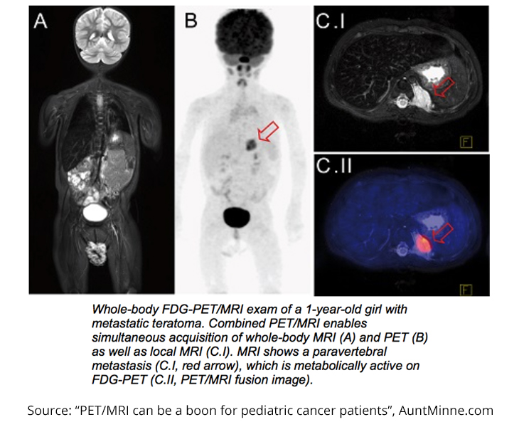
 

Pediatric Oncology Patients to Benefit from FDG-PET/MRI: Safer Option Than FDG-PET/CT

A recent study led by Dr. Sergios Gatidis of the University of Tübingen revealed an easier – and potentially safer – method of identifying malignancies in pediatric cancer patients. Gatidis and his colleagues pursued this research to improve the quality of care for younger patients. Similar studies have been performed for juvenile and adult oncology patients, but the specific challenges of younger patients set this study apart.

Not only do tumors act differently depending on the age of the patient, but also most children have a difficult time sitting still for extended periods of time, rendering some FDG-PET/CT scans unreadable. The problem then becomes needing to sedate patients, exposing them to more radiation and more medication than what is actually required.

telerad-sept-blog

This study included nine patients – six boys and three girls – between the ages 1 and 6. All the patients were scheduled to have FDG-PET/CT scans for diagnostic purposes before participating in the study. Two different teams of readers analyzed the images, and the FDG-PET/MRI scans gave more certain diagnostic results than the PET/CT scans. Overall, six of the seven scans that were read differently by PET/CT and PET/MRI had a “clearer morphologic correlate in MRI compared to CT.” Furthermore, 5 of 10 MRI scans led to changes in patient management.

The results of the study show that the MRI scans were just as good as CT scans, if not better. By changing the imaging process from PET/CT to PET/MRI, pediatric patients will hopefully be able to experience less total examination time, less exposure to harmful radiation and sedatives, and more accurate treatment planning.

Brain MRI Interpretation Detects Potential Stroke, Death with 3mm Lesions in Asymptomatic Patients

MRI brain interpretation

On our teleradiology blog, we like to regularly post articles on findings that have been discovered in the medical field, especially when they relate to key areas of our business.

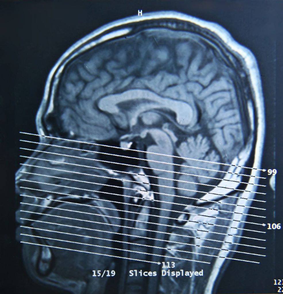
With that in mind, we found the following study on the possibilities for MRI brain interpretations fascinating, and we look forward to following any developments in the future. Give this Specialty Teleradiology summation on MRI brain scans a look.

MRI Brain Interpretations and Their Role in Stroke Prediction

According to a recent study published in the Annals of Internal Medicine it was reported that patients with one or two small lesions were five times more likely to have a stroke and/or have a stroke-related death. Researchers from the University of Mississippi Medical Center, along with a few other U.S. institutions, conducted the study.

Here are some of the key highlights about the conduction of the study:

  • Researchers examined data from 1,884 patients from Forsyth County, North Carolina, and Jackson, Mississippi, who had previously participated in the Atherosclerosis Risk in Communities (ARIC) study.
  • Patients “received scans on a 1.5-tesla MRI system with an imaging protocol that included T1-weighted, T2-weighted, and proton density-weighted imaging of the whole brain.”
  • The data showed that the hazard ratio of stroke-related death for patients with any sized lesion was 6.97, compared to the hazard ration of 1.89 for all-cause death patients.

As one of the first studies to examine such data, the results have indicated the importance of further examination of similar situations. Small lesions could also indicate a future risk for other cerebrovascular diseases, such as dementia and other mobility problems. As medical imaging such as MRI brain interpretation becomes more sophisticated, we can potentially understand the causes and/or risks of 3 mm lesions and what they could mean for visibly asymptomatic patients with brain MRIs.

Further Industry Insights

We cover a lot of material on our blog, featuring information about regional imaging centers, useful media resources, historical perspectives on procedures, and more. Here are a few select posts to help you discover more about the radiology field:

You can additionally subscribe to our newsletter to get monthly updates from us.

Contact Us

Interested in our teleradiology services like MRI brain interpretations? Contact our team today!

Improving Treatment for Patients with Metastatic Spine Disease Through Assessment Agreement

In an effort to improve treatment for skeletal metastatic cancer – the most deadly form of the cancer – the Spanish Back Pain Research Network, Kovacs Foundation, conducted a study to see correlation between the Tomita and modified Bauer scores when determining the prognosis and surgical strategy. The goal of the study was to establish a standardized method of communicating the severity of metastatic spine disease through medical oncologists, radiologists, radiation oncologists, orthopedic surgeons, and neurosurgeons.

Eighty-three doctors with multiple levels of post-residency practice, specialties, and hospital complexity participated in the study and the first 90 patients that met the inclusion criteria and did not meet any of the exclusion criteria were accepted as subject participants. Intraobserver and interobserver variability were taken into account amongst the clinicians. Patient information was limited to two (2) MRIs and a clinical vignette of basic patient history and treatment.

Both intra- and inter-observer agreements were almost entirely “almost perfect” and “substantial.” This high agreement correlation could improve the efficiency and efficacy of treatment for patients with metastatic spine diseases. Previous studies have examined the benefits one scoring system over the other, but this is one of the first studies to utilize both scores for a better inter-disciplinary communication system. Future studies may have a more specific group of doctors, particular cancer subtypes and/or location of metastases, or hospital categories to further determine the best scenarios for interobserver communication.

Detecting Enthesitis in Patients with Psoriatic Arthritis and Axial Spondyloarthritis with WBMRI

mri detecting enthesitis

Relatively new to the diagnostic imaging world is the use of whole body magenetic resonance imaging (WBMRI). Just as the name suggests, this MRI is taken from the patient’s head to toes. While the image is comprehensive, the image resolution is lower than with standard MRIs. Even so, a recent study from the Copenhagen Center for Arthritis Research gave preliminary indication that WBMRIs could help with detecting enthesitis in arthritic patients.

The study was performed on 36 patients with psoriatic arthritis (PsA) or axial spondyloarthritist (axSpA), as well as a control group of 12 healthy patients. Patients underwent a physical examination and an imaging session that included contrast enhancement and coronal slice orientation for spine, hip, knee and ankle, sagittal orientation for the neck, and axial orientation for the feet. One study summary identified that part of the study included determining whether or not the WBMRIs were “readable” or “not readable.” Readable enthesitis images were defined as “the presence of bone marrow edema, soft tissue edema, change in tendon thickness, erosions or enthesophytes in adjacent bones, and fluid in or around tendons or adjacent bursae.”

The study’s results showed that WBMRIs did indicate enthesitis in the PsA and axSpA patients more than the healthy patients and that enthesitis was identified in 17% of the readable sites through WBMRIs and clinical enthesitis was present in 22% of the readable sites. This study was the first of its kind, but has given hope to doctors that WBMRIs could help rule out other inflammatory diseases and perhaps help doctors with diagnoses and treatment plans.

If more studies are performed, a larger sample would be used and cost-effectiveness would become more of a consideration.