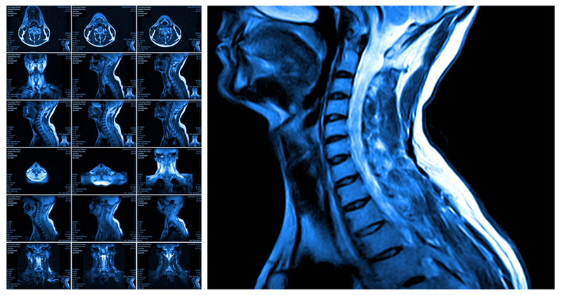
As part of our dedication to making our radiology blog a source for key insights on diverse topics within the medical field, for this post we decided to focus on the ways MRI spine interpretations are playing a role in advancing patient care.
So if you’re interested in why lumbar and cervical spine MRI results interpretations are so essential, read on to discover recent findings.
MRI Spine Interpretations for Ankylosing Spinal Disorders
According to a study published in The Spine Journal, patients with ankylosing spinal disorders who have non-ankylosed levels can benefit from MRI spine interpretations in a diagnostic role. Let’s take a look at a few key insights:
- MRI had previously been proposed as a routine tactic to aid in the identification of noncontiguous fractures in patients, and those who suffer from back and neck pain due to an injury or from an unknown source.
- For the study, the researchers sought to measure how often an MRI would be able to identify an injury that had not previously been identified through CT scans.
- With 124 patients utilized for the study, MRI interpretations for the spine were able to reveal injuries in 4.8% that had not previously been diagnosed via CT scans. This new information brought about alterations, both surgical and nonsurgical, in the patients’ treatment plans.
The Next Steps Following Incidental Renal Lesions Found During Lumbar Spine MRI Interpretations
In a study published in the American Journal of Roentgenology, and reported by Radiology Business, the role of follow-up imaging after the discovery of incidental renal lesions was examined. Some of the key points:
- 149 renal lesions that had been identified from spine MRI interpretations were studied by imaging specialists.
- Of these 149 renal lesions, 115 were basic cysts while 34 were complex renal lesions.
- The complex renal lesions were detected by the readers at 94%, and these same readers also identified all malignant and potentially malignant lesions.
- Due to these findings, the authors argued that follow-up renal imaging may not be necessary for many cases of incidentally discovered renal lesions.
Spinal Muscular Atrophy Disease May Be Uncoverable from MRIs
According to a case report from Clinical Imaging, MRI spine interpretations may be able to detect the presence of spinal muscular atrophy (SMA) as well as monitor the disease itself. Some key findings:
- According to the report, imaging was able to identify indications of ventral nerve atrophy in the lower back of a child who had SMA type 2. The report notes that ventral nerve atrophy is usually only discovered during postmortem examinations.
- The patient in question had had difficulty latching during breastfeeding and had developed poor muscle tone for his age, though brain MRIs showed no abnormalities. The spinal MRI, however, detected SMA.
- Moving forward, the authors of the report recommend analyzing further spine MRI data to test the consistency of these findings and determine whether MRIs can be an effective non-invasive biomarker for monitoring disease progression.
Follow Our Radiology Blog
For more insights on lumbar and cervical spine MRI interpretations, PET/CT reporting, imaging centers, radiology resources and more, be sure to follow our blog. And if you’re in need of reliable teleradiology services with quick turnaround, contact our representatives today!
