PET Scans Detect Early Signs of Alzheimer’s

Picture1

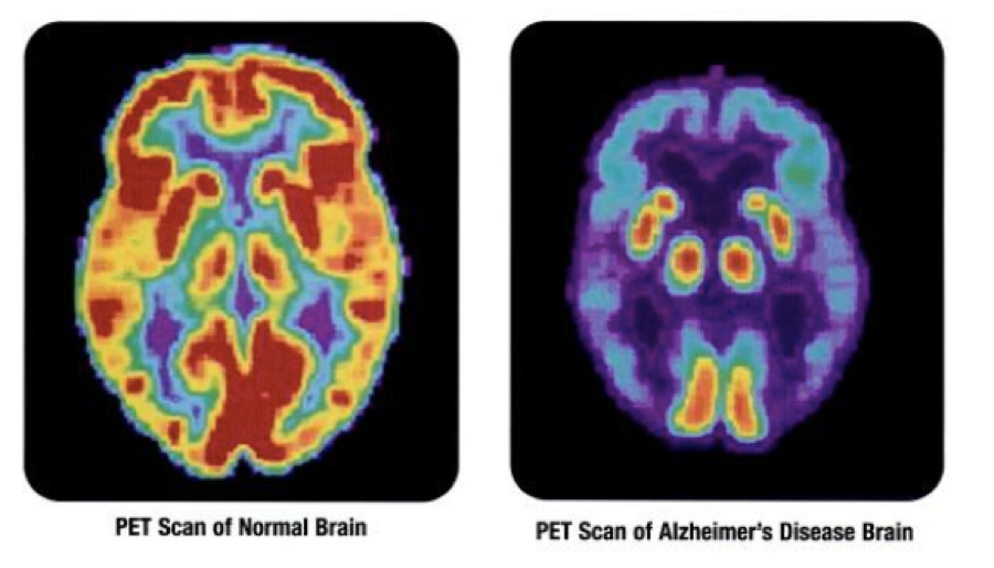
For the first time, scientists have shown that PET scans can identify the progressive stages of Alzheimer’s. A recent study performed at the University of California, Berkeley and published in the journal “Neuron” shines new light on a disease affecting millions of Americans. The scans were able to detect the progression of the disease before patients showed any other symptoms. The research could pave the way for improved Alzheimer’s diagnostic protocol.

To better understand the progression of Alzheimer’s, researchers took PET scans of 53 adults between the ages of 20 and 90. Some participants showed signs of Alzheimer’s, while others did not. Breakthroughs in PET scan technology have given doctors more powerful tools to see abnormalities that wouldn’t have been visible before. Previously, the clearest scans were limited to people who had already died. Using modern PET scans, the team observed varying stages of Alzheimer’s progression.

The researchers analyzed scan results and searched for accumulated tau and beta-amyloid proteins, as they have previously been associated with the disease. The results showed that once tau proteins had spread beyond the medial temporal lobe, outward symptoms of Alzheimer’s were present. Amyloid proteins also appear to play a role in this process. The team acknowledged that more research is necessary to utilize PET scans as an Alzheimer’s diagnostic tool. Regardless, the results are a promising indication that doctors can identify the disease in its early stages.

Scans Predict Transplant Survival Rates

scan


Radiology recently published a study that shows FDG-PET/CT scans can predict survival rates for lymphoma patients following stem cell transplantations. The presence of FDG-avid lesions on scan results was a strong indicator of poor outcomes. Patients with lesions who received allogeneic stem cell transplantation were half as likely to survive as those who tested negative. For patients who received autologous stem cell transplantation, the difference was even clearer. The presence of FDG-avid lesions led to a two-year progression-free survival rate below 20%. Patients without FDG-avid lesions were four times more likely to survive. The study suggests that doctors should account for scan results before moving forward with a stem cell transplant.

To conduct the study, researchers retrospectively analyzed scans from 73 allogeneic transplant patients and 102 autologous transplant patients. These patients had been administered 12 mCi to 15 mCi of FDG before their PET/CT scans. The researchers only examined scans that were completed 3 months prior to a transplant. They noted any FDG-avid lesions and the maximum standardized uptake value for the most active lesion. The doctors categorized any scan with an FDG-avid lesion as PET-positive and those without as PET-negative.

The correlation between high FDG avidity and low survival rates gives doctors a better idea of what results to expect following a stem cell transplant. Some physicians already use scan results as a guide when deciding whether to move forward with the expensive procedure. The study’s results suggest that multiple rounds of chemotherapy may be necessary before a patient undergoes an allogeneic or autologous transplant. It’s clear that scans play an important role in a patient’s prognosis. Stem cell transplants are much more likely to be successful when doctors can identify the right candidates.


Immunotherapy and Scan Analysis

Lung scan

Immunotherapy has been a growing area of research in the field of lung cancer care. As the treatment gains popularity, doctors are learning more about how it differs from traditional chemotherapy. In the past, scans that showed growing disease areas and new lesions after therapy indicated cancer progression. This remains true for treatments such as standard chemo, Tarceva and Avastin. According to Lungevity, scans for patients who receive immunotherapy sometimes show growing lesions even when the patient feels fine and their cancer symptoms are improving.

Researchers have found that new or growing lesions that result from immunotherapy aren’t necessarily full of cancer cells. Instead, the host’s immune cells tend to group in these areas to attack the cancer cells. Eventually the lesion disappears completely with further immunotherapy. Immune cells sometimes attack tumor cells that were too small to see in previous scans.

Patients undergoing clinical trials of immunotherapy have provided a wealth of information to researchers. Previous ideas of what a good scan looks like are quickly becoming obsolete. The new research suggests that a biopsy may be necessary when a scan indicates new or growing lesions in immunotherapy patients. When the clinical picture looks good, but the lesions look bad, discontinuing immunotherapy may not be the best course of action.

The High Accuracy of Heart CT Scans

John Hopkins researchers have shown that non-invasive CT angiograms are more effective at detecting clogged coronary arteries than exercise stress tests. Around 15 million Americans visit physicians each year with symptoms of a clogged artery. Chest pain, shortness of breath and fatigue often warrant further testing to spot any blockages. Cardiologists typically rely on a stress test that measures blood flow after a patient walks on a treadmill. American Heart Association guidelines currently call for this test when patients show signs of coronary artery disease. The new study indicates that CT angiograms may be a better testing method due to lower radiation levels, higher accuracy and comparable cost.

The gold standard for detecting clogged arteries remains an invasive cardiac angiography. This test involves threading a catheter into a patient’s heart blood vessels. It’s a process that carries certain risks, so cardiologists utilize an initial “gatekeeper” test to identify the people most likely to benefit from it. The John Hopkins study shows that stress tests only correctly identify blockages in 6 out of 10 people. Comparatively, CT scans accurately spotted blockages in 9 out of 10 patients.

Participants of the study underwent all three testing methods, providing a direct comparison of the effectiveness of each. For CT angiograms, clinicians inject dye into a patient’s circulation to visualize the arteries. The test spots blockages caused by fatty buildups or clots in the interior of arteries. Clinicians have shied away from this process in the past due to limited evidence of its efficacy. The recent study examined 391 patients in 16 hospitals and eight countries. It was the largest study of its kind to date.

The researchers hope that the results show clinicians that CT angiograms are a more accurate alternative than the widely used stress tests. Both tests provide a first-line diagnosis before an invasive cardiac angiography. The unprecedented scale of the study gives cardiologists solid evidence to support CT scanning. For patients concerned about blocked arteries, the results are promising. A near-perfect diagnosis rate seems more realistic than ever before.

Advantages of an Abbreviated Breast MRI Protocol

telerad pic 2


A recent study has found evidence that a shorter noncontrast-enhanced breast MRI protocol could reduce scanning times and improve information gathering for suspicious lesions. The study was performed by the German Cancer Research Center and led by Dr. Sebastian Bickelhaupt. The protocol, referred to as diffusion-weighted imaging with background suppression (DWIBS), had a negative predictive value of 92%, comparable to conventional diagnostic MRIs. The DWIBS method was able to obtain images in just seven minutes vs. 30 minutes for the standard protocol. The approach may increase the rate of positive biopsies.

MRI exams are often used to complement conventional mammography, which has a high false-positive rate. Traditional MRI exams use contrast and can be time-consuming. The authors were interested in abbreviated exams due to recent papers demonstrating the benefits of such an approach. They compared this method with an abbreviated contrast-enhanced protocol as well as a full diagnostic breast MRI.

The study took place between September 2014 and January 2015. The researchers enrolled 50 women who had suspicious screening mammogram results and an indication for biopsy. They performed the exams on a 1.5-tesla scanner. The study was performed between two imaging sites with two readers blinded to the each other’s data and interpretations.

The DWIBS method came out positive for 100% of the histopathologically confirmed invasive breast cancers among the test group. The only exceptions were two lesions with pure microcalcifications. The authors noted that these are “notoriously invisible” when using MRI. There was no significant difference between the accuracy of full diagnostic and the abbreviated protocols.

The study had some limitations, such as a patient group with a greater probability of malignant lesions than an unselected population, which the researchers acknowledged. The team concluded that the DWIBS mammography protocol was accurate in ruling out malignancy after an initial mammogram screening.