Diagnosing Appendicitis with MRIs

diagnose-appendicitis-with-mris
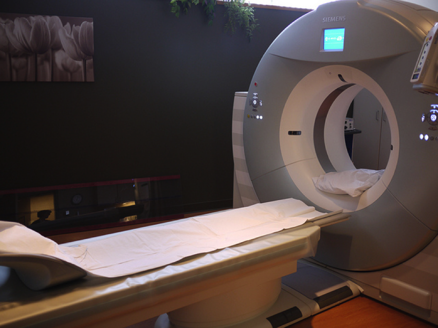
Children and young adults with right lower quadrant (RLQ) pain may benefit from an MRI diagnosis, according to a new study published in Radiology. A University of Arizona team determined the accuracy of unenhanced MR imaging for patients in the retrospective study. In all, the researchers examined imaging from 403 patients with an age range of 3-49 who experienced RLQ pain and received an MRI. The team suspected that intravenous or oral contrast methods were unnecessary with the use of an MRI diagnosis.

To learn more about MRI accuracy, the team looked at clinical records that documented MRI room time as well as prospective image interpretations. While some of the patients suffered from appendicitis, others were experiencing other types of abdominal pain. Attaining information for patient outcomes was essential for the study. The team was able to collect final results for patients with documentation including surgical results, telephone follow-ups, medical records and expert panel assessments.

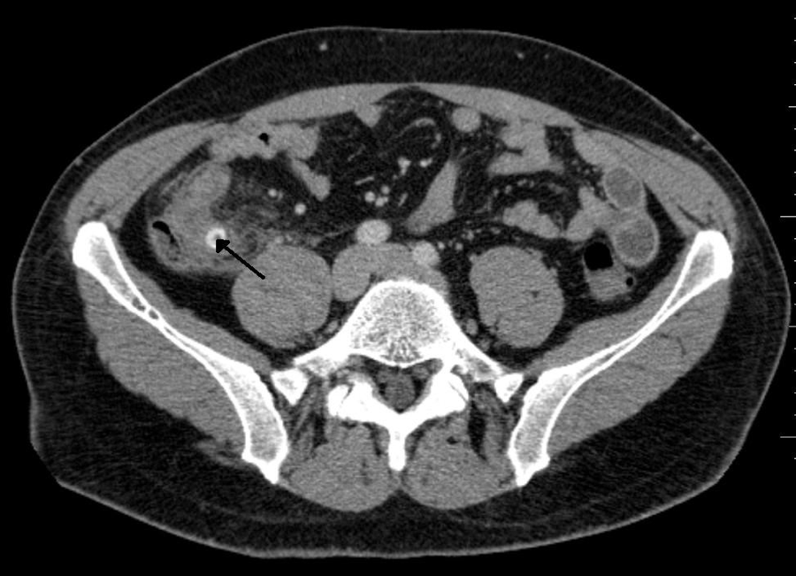
Gathering patient data was important, but the results mattered most. The study revealed that the average MRI room time was 14 minutes. Among the 403 patients, 67 tested positive for acute appendicitis, while 336 had negative results. The MRIs had 97% sensitivity and 99.4% specificity. 51.5% of the patients were diagnosed with an illness other than appendicitis. This high degree of accuracy led the researchers to conclude that MRIs are an effective way to evaluate and diagnose RLQ pain.

Mobile App Helps Doctors Choose An Imaging Test

doctor-clipboard


For primary care practitioners, choosing the right imaging test in response to a patient’s symptoms can be difficult. Resident MD Winnie Tsao of the University at Buffalo wants to help doctors determine the appropriate test for a variety of medical situations. She developed a mobile application named Radiology Consult that provides an imaging test recommendation for a faster and more cost-effective diagnosis. In just a few taps, physicians can uncover the appropriate course of action for their patients, without the need to page through huge textbooks.

The app uses complex algorithms to identify common medical conditions, and the most relevant imaging test for each disease or injury. In addition to a recommendation, the app lists procedural preparations and a description of the advantages and drawbacks for each test. Interacting with the app is simple, as it uses straightforward language and a simple step-by-step process. It should be noted that while the app helps doctors choose an imaging test, it does not interpret results or recommend a diagnosis ­– that’s something only a skilled radiologist can do. The app was built for Apple’s App Store, but Tsao plans to create an Android version in the future.

Tsao’s app benefits doctors, patients and even health insurance providers, primarily by preventing doctors from performing unnecessary – and costly- tests. Since Radiology Consult works right at point of care, doctors cut a significant amount of time from the diagnostic process. The app has received favorable reviews, and Tsao continues to work on development during her residency. Radiology Consult has gained popularity due to its convenience and originality. Tsao discovered that practitioners wanted an easier way to choose imaging tests, so she built an effective solution from the ground up. It’s just one more tool for doctors to use, and it’s already in their pockets.

CT Scan Accuracy in Detecting Lung Cancer

lungs

A recent study published in The Lancet Oncology journal suggests that it only takes one CT scan to eliminate the possibility of lung cancer among high-risk patients. This contradicts the current recommendation that smokers receive annual tomography screenings to detect early signs of the disease. CT scans outperform x-rays when it comes to detecting lesions, but also emit more radiation. If the results of a single CT scan show no signs of cancer, patients may be able to avoid unnecessary health care costs with a yearly screening.

To conduct the study, researchers retrospectively analyzed CT scan results from over 26,000 smokers and ex-smokers. Around 19,000 of the patients tested negative for signs of lung cancer. The team then looked at scans from the same group, conducted one year later. The incidence of lung cancer among patients who initially showed no irregularities was extremely low. The researchers concluded that the second scan may not have been necessary considering the downsides that come with frequent scanning.

One potential outcome from the study is that doctors may choose to conduct high-accuracy CT scans less frequently for lung cancer screenings. Because the study was based on retrospective analysis, more research is needed before anyone draws concrete conclusions. Additionally, the ideal interval between CT scans is still unclear. The study is sure to inspire continued research in the area of lung cancer scans.

Scans Predict Transplant Survival Rates

scan


Radiology recently published a study that shows FDG-PET/CT scans can predict survival rates for lymphoma patients following stem cell transplantations. The presence of FDG-avid lesions on scan results was a strong indicator of poor outcomes. Patients with lesions who received allogeneic stem cell transplantation were half as likely to survive as those who tested negative. For patients who received autologous stem cell transplantation, the difference was even clearer. The presence of FDG-avid lesions led to a two-year progression-free survival rate below 20%. Patients without FDG-avid lesions were four times more likely to survive. The study suggests that doctors should account for scan results before moving forward with a stem cell transplant.

To conduct the study, researchers retrospectively analyzed scans from 73 allogeneic transplant patients and 102 autologous transplant patients. These patients had been administered 12 mCi to 15 mCi of FDG before their PET/CT scans. The researchers only examined scans that were completed 3 months prior to a transplant. They noted any FDG-avid lesions and the maximum standardized uptake value for the most active lesion. The doctors categorized any scan with an FDG-avid lesion as PET-positive and those without as PET-negative.

The correlation between high FDG avidity and low survival rates gives doctors a better idea of what results to expect following a stem cell transplant. Some physicians already use scan results as a guide when deciding whether to move forward with the expensive procedure. The study’s results suggest that multiple rounds of chemotherapy may be necessary before a patient undergoes an allogeneic or autologous transplant. It’s clear that scans play an important role in a patient’s prognosis. Stem cell transplants are much more likely to be successful when doctors can identify the right candidates.


Immunotherapy and Scan Analysis

Lung scan

Immunotherapy has been a growing area of research in the field of lung cancer care. As the treatment gains popularity, doctors are learning more about how it differs from traditional chemotherapy. In the past, scans that showed growing disease areas and new lesions after therapy indicated cancer progression. This remains true for treatments such as standard chemo, Tarceva and Avastin. According to Lungevity, scans for patients who receive immunotherapy sometimes show growing lesions even when the patient feels fine and their cancer symptoms are improving.

Researchers have found that new or growing lesions that result from immunotherapy aren’t necessarily full of cancer cells. Instead, the host’s immune cells tend to group in these areas to attack the cancer cells. Eventually the lesion disappears completely with further immunotherapy. Immune cells sometimes attack tumor cells that were too small to see in previous scans.

Patients undergoing clinical trials of immunotherapy have provided a wealth of information to researchers. Previous ideas of what a good scan looks like are quickly becoming obsolete. The new research suggests that a biopsy may be necessary when a scan indicates new or growing lesions in immunotherapy patients. When the clinical picture looks good, but the lesions look bad, discontinuing immunotherapy may not be the best course of action.